Research
Magnetic resonance imaging biomarkers enable a comprehensive characterization of tissue providing functional, physiological, metabolic, cellular and molecular information beyond anatomical structures.
In recent years, hyperpolarization techniques allowed to increase the inherent low sensitivity of magnetic resonance by more than four orders of magnitude, opening new applications ranging from the tracking of chemical reactions in real-time to the direct monitoring of metabolic processes in vivo.
Another functional MRI technique that has already shown promise in characterizing tissue in clinical studies is diffusion-weighted MRI (DW-MRI). It enables the quantitative assessment of the apparent diffusion coefficient (ADC) that depends on microstructural tissue properties, including cellularity and intercellular matrix composition.
In our group, we focus on the development of sensitive hyperpolarized molecules for imaging metabolism and pH as well as advanced diffusion MRI techniques aiming to provide novel imaging biomarkers to characterize tissue function. We exploit a multimodal approach utilizing the combined strength of high sensitivity PET tracers for simultaneous PET/MRI. We develop fundamentally new methods across all scales from in vitro to in vivo to patients.
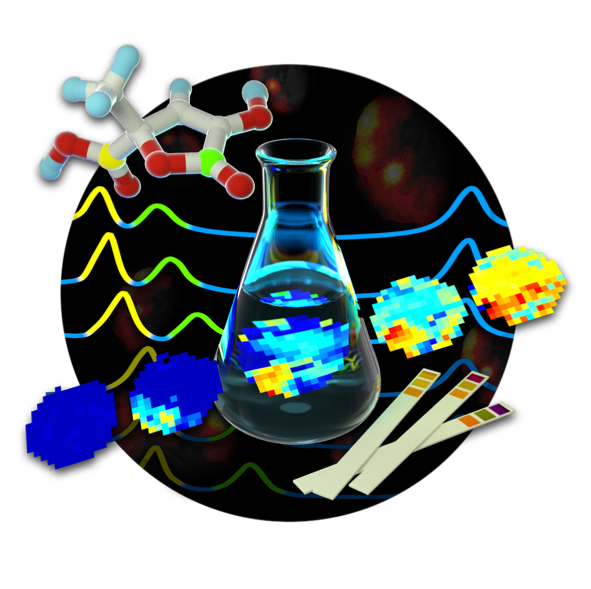
pH Biosensors
Resolving spatiotemporal pH changes non-invasively in vivo using hyperpolarized pH sensor compounds.
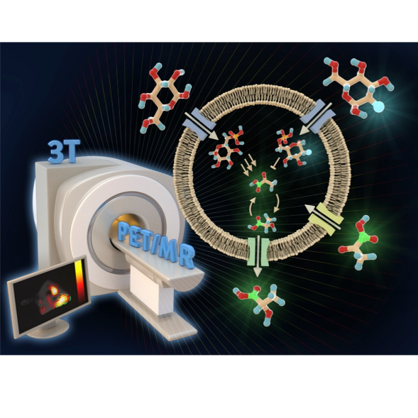
Metabolic Imaging
Quantifying metabolism in vivo using PET/MRI with hyperpolarized compounds and development of MRI pulse sequences.
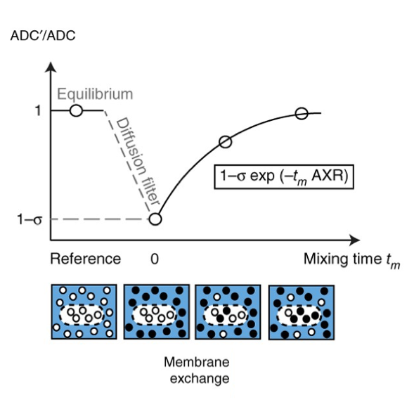
Diffusion and Exchange
Characterizing tissue properties – including cellularity and water permeability – with novel MRI pulse sequences.


