Latest News
-
 Inside Front Cover in Advanced ScienceApril 23, 2026/0 Comments
 -
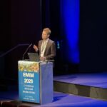 European Molecular Imaging Meeting 2026March 26, 2026/
 -
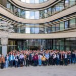 THINC Conference Munich 2026March 23, 2026/
 -
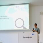
 PhD Defense of Wolfgang GottwaldMarch 3, 2026/
 -
 Munich Metabolic Methods MeetingJanuary 20, 2026/
 -
 MRI Christmas 2025December 24, 2025/
 -
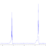
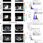 New Paper: Early Detection of Cell Death Using MRIDecember 22, 2025/
 -
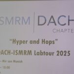 Hyper&Hops MeetingJune 26, 2025/
 -
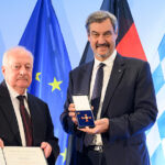 Bundesverdienstkreuz for Prof. Axel HaaseJune 25, 2025/
 -
 12th Munich Cancer RetreatJune 24, 2025/