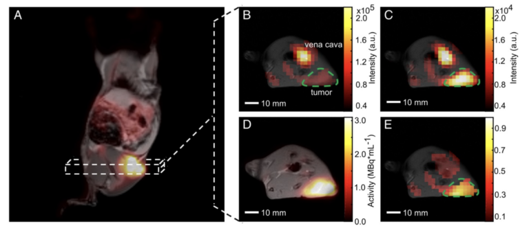
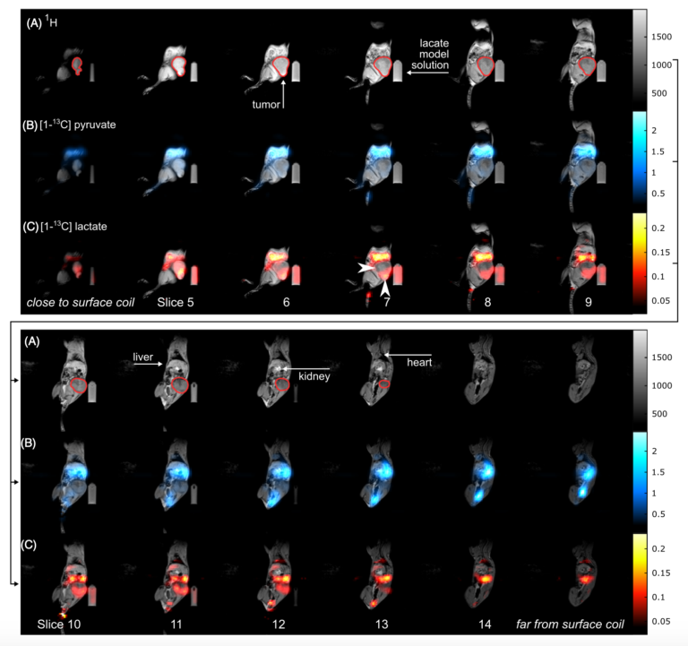
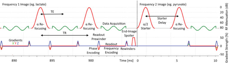
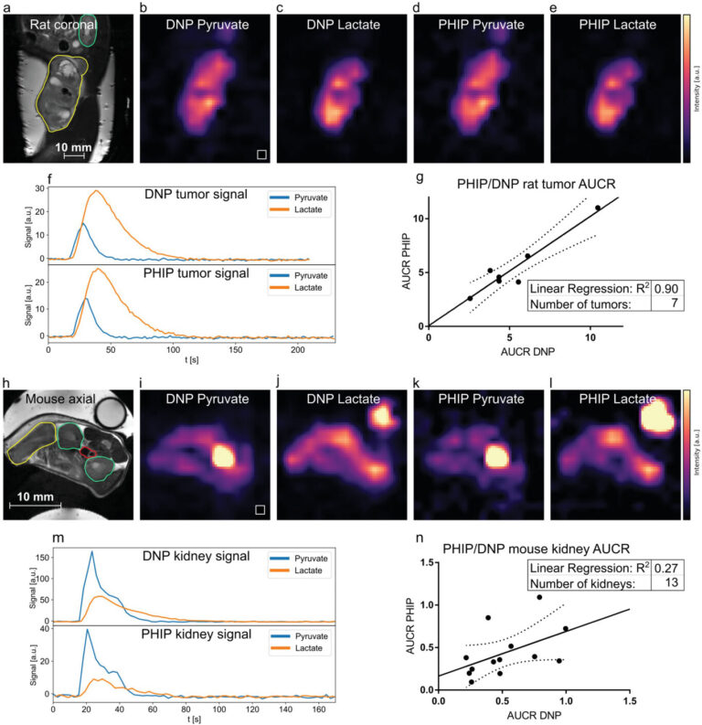
Hyperpolarized 13C MRI provides a unique insight into metabolism and tissue function. It utilizes molecular transformations as a mechanism for imaging contrast. The most successful hyperpolarized 13C MRI probe so far is [1-13C]pyruvate. Pyruvate is a key metabolite in energy metabolism and, as the end product of glycolysis, it is further metabolized to lactate, alanine or to acetyl-CoA (after cleavage of 13CO2), which then enters the tricarboxylic acid (TCA) cycle. By quantifying pyruvate’s downstream metabolism, glycolytic tissue can be differentiated from tissue dominated by aerobic respiration. Besides dissolution dynamic nuclear polarization (dDNP), which operates at liquid helium temperature close to absolute zero temperature, other methods for generating hyperpolarized 13C-labeled probes are based on polarization transfer from parahydrogen and require substantially less technological efforts and cost. In a recent collaborative effort, we have demonstrated preclinical evidence for safe and reliable polarization and in vivo administration of [1-13C]pyruvate using parahydrogen (See Figure 4, Nagel et al. 2023). We aim to further translate this approach to the clinic with first experiments in human cancer patients planned in 2026 at the TUM University Hospital.
 Inside Front Cover in Advanced ScienceApril 23, 2026/0 Comments
 European Molecular Imaging Meeting 2026March 26, 2026/
 THINC Conference Munich 2026March 23, 2026/
 PhD Defense of Wolfgang GottwaldMarch 3, 2026/
 Munich Metabolic Methods MeetingJanuary 20, 2026/
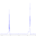 MRI Christmas 2025December 24, 2025/
 New Paper: Early Detection of Cell Death Using MRIDecember 22, 2025/
 Hyper&Hops MeetingJune 26, 2025/
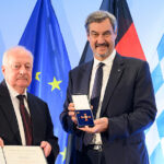 Bundesverdienstkreuz for Prof. Axel HaaseJune 25, 2025/
 12th Munich Cancer RetreatJune 24, 2025/