Congratulations to Elisabeth Bliemsrieder and coauthors on the publication of ‘Hyperpolarized 13C pyruvate magnetic resonance spectroscopy for in vivo metabolic phenotyping of rat HCC’ in Scientific Reports.
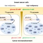
The in vivo assessment of tissue metabolism represents a novel strategy for the evaluation of oncologic disease. Hepatocellular carcinoma (HCC) is a high-prevalence, high-mortality tumor entity often discovered at a late stage. Recent evidence indicates that survival differences depend on metabolic alterations in tumor tissue, with particular focus on glucose metabolism and lactate production. Here, we present an in vivo imaging technique for metabolic tumor phenotyping in rat models of HCC. Endogenous HCC was induced in Wistar rats by oral diethyl-nitrosamine administration. Peak lactate-to-alanine signal ratios (L/A) were assessed with hyperpolarized magnetic resonance spectroscopic imaging (HPMRSI) after [1-13C]pyruvate injection. Cell lines were derived from a subset of primary tumors, re-implanted in nude rats, and assessed in vivo with dynamic hyperpolarized magnetic resonance spectroscopy (HPMRS) after [1-13C]pyruvate injection and kinetic modelling of pyruvate metabolism, taking into account systemic lactate production and recirculation. For ex vivo validation, enzyme activity and metabolite concentrations were spectroscopically quantified in cell and tumor tissue extracts. Mean peak L/A was higher in endogenous HCC compared to non-tumorous tissue. Dynamic HPMRS revealed higher pyruvate-to-lactate conversion rates (kpl) and lactate signal in subcutaneous tumors derived from high L/A tumor cells, consistent with ex vivo measurements of higher lactate dehydrogenase (LDH) levels in these cells. In conclusion, HPMRS and HPMRSI reveal distinct tumor phenotypes corresponding to differences in glycolytic metabolism in HCC tumor tissue.
Download the open access article here.
Bliemsrieder, Elisabeth, et al. “Hyperpolarized 13 C pyruvate magnetic resonance spectroscopy for in vivo metabolic phenotyping of rat HCC.” Scientific reports 11.1 (2021): 1-9.

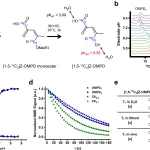
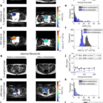
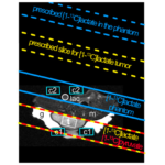
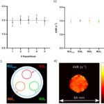
Normalized time-intensity curves of subcutaneous tumors derived from LA-L and LA-H cell lines. [1-13C]lactate and [1-13C]pyruvate signal curves of LA-L (a, c) and LA-H (b, d) tumors before (a, b) and after (c, d) application of the kinetic-model-derived scaling factors to the [1-13C]lactate signal curves. Metabolite intensities are normalized to pyruvate peak intensity. kpl-values of the same LA-L and LA-H tumors (e) and corresponding LDH activity of their extracts (f). Both Student’s T-test p < 0.001.


![Parahydrogen-Polarized [1-13C]Pyruvate for Reliable and Fast Preclinical Metabolic Magnetic Resonance Imaging](https://schillinglab.com/wp-content/uploads/2023/09/advs6326-fig-0001-m-150x150.jpg)